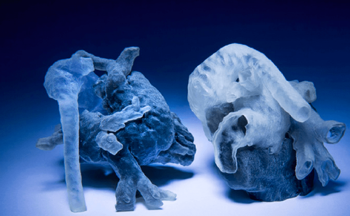
The truly revolutionary aspect of this 3D-printed model heart is the high degree of detail and personalization it gives surgeons preparing for a procedure. While generic models of the heart have existed for years, most patients requiring heart surgery don’t have generic hearts — indeed, the irregularities found in their organs necessitate surgery, and as such, doctors need to have as specific of information as possible to ensure success.
When examining an MRI, doctors are given access to cross sections of a 3D object, like the heart. Cross sections are black and white, with the boundaries between the regions of different colors serving as a potential indicator of some anatomical structure, like a chamber or a blood vessel. The key in creating a model of the heart is figuring where these boundaries are and what they mean — a process known as “image segmentation.”
Historically, scientists and doctors manually performed this task, carefully poring over MRI scans to determine where boundaries are. But with high-precision scans, this means going through some 200 cross sections, which simply isn’t feasible in a short time frame. Explained Golland, “[Doctors] want to bring the kids in for scanning and spend probably a day or two doing planning of how exactly they’re going to operate. If it takes another day just to process the images, it becomes unwieldy.”
But Golland’s work solves for this problem in a novel way. Rather than having a human establish where all the boundaries were in all 200 cross sections, she and her team simply had a doctor look at a few sections, and then developed an algorithm that could do the rest. They found that after a live expert identified just one-ninth of the total area, they could then automate away the remaining cross sections with 90 percent accuracy. Pretty amazing when efficiency is taken into consideration.
“I think that if somebody told me that I could segment the whole heart from eight slices out of 200, I would not have believed them,” Golland told MIT news. “It was a surprise to us.”
Already, doctors are thrilled by the implications of this work. “Absolutely, a 3D model would indeed help,” Sitaram Emani, a cardiac surgeon at Boston Children’s Hospital told the media. “We have used this type of model in a few patients, and in fact performed ‘virtual surgery’ on the heart to simulate real conditions. Doing this really helped with the real surgery in terms of reducing the amount of time spent examining the heart and performing the repair.”
Moreover, he noted, having an actual model will make explaining complex situations to concerned family members much simpler. Said Emani, “This gives them a better visual, and many patients and families have commented on how this empowers them to understand their condition better.”
And of course, it’s always great to practice a bit before the real thing — especially when that real thing is open-heart surgery.


